Ultrasonography examples
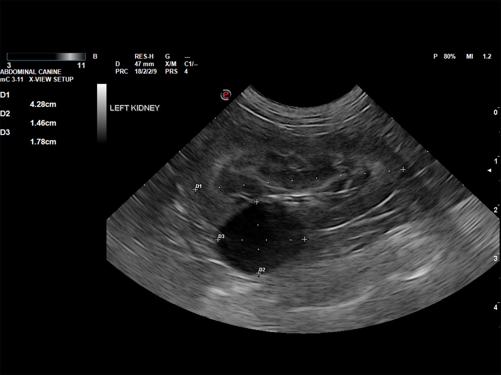
Longitudinal view of a canine kidney with a large anechoic cyst arising from the cortex and expanding the capsule. Small renal cysts can be commonly seen particular in older patient’s kidneys and are usually benign unless they become very large and then can be a source of pain and discomfort.

Transverse view of the liver with the gallbladder located within the right lobar region (hypoechoic black structure on the left in the picture). This liver preserved normal echogenicity, echotexture and architecture.
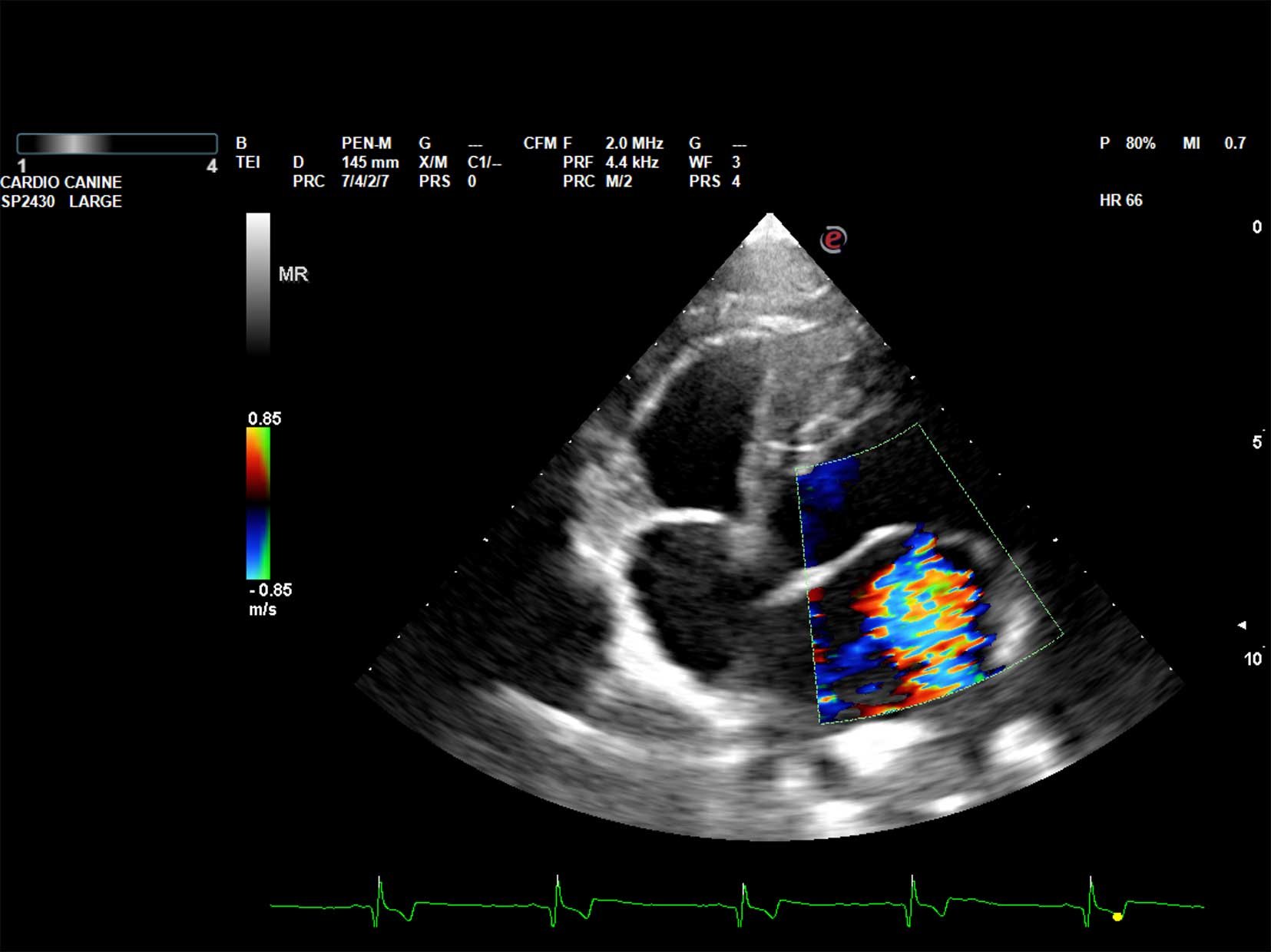
Left side apical view of the heart with Colour Doppler showing moderate to severe mitral regurgitation (blood flow into the left atrium when the mitral valve is closed). This can be seen with canine myxomatous mitral valve disease (MMVD) which is commonly diagnosed in breeds such as the Cavalier King Charles Spaniel, however, other breeds can also develop this disease.

Two hyperechoic rounded structures casting strong distal acoustic shadowing (the black band underneath each structure) located in the dependent portion of the urinary bladder lumen. These structures are consistent with uroliths (bladder stones) and in a feline (cat), they are most likely struvite uroliths.